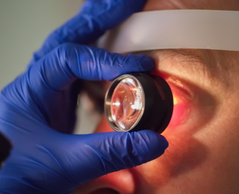
This test involves photographing the back of an eye, also known as the fundus. Specialized fundus cameras consist of an intricate microscope attached to a flash-enabled camera.
Fundus photography is a specialized imaging technique used to capture detailed photographs of the interior surface of the eye, particularly the retina, optic disc, and macula. This type of photography helps diagnose and monitor various eye conditions, such as diabetic retinopathy, glaucoma, and age-related macular degeneration. The images obtained can provide valuable insights into the health of the eye and can aid healthcare professionals in tracking disease progression or treatment effectiveness.


